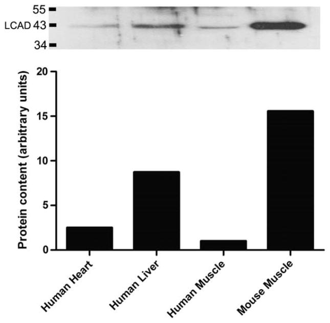
Fig. 3.
LCAD protein content in enriched mitochondrial samples. LCAD protein is detectable in 10 μg of human isolated mitochondria from heart and liver homogenate (lanes 1&2). Thirty μg of isolated mitochondria from human skeletal muscle was required to quantify LCAD expression (lane 3). Thirty μg of isolated mitochondria from mouse quadriceps muscle was loaded as a comparison (lane 4). Graph: The quantified bands were normalized to ponseau (total protein). LCAD expression in human mitochondrial enriched samples was most abundant in human liver. Human skeletal muscle had the least amount of LCAD protein which was 16 times less that of mouse skeletal muscle. Comparative analysis, N=1 for all samples.

