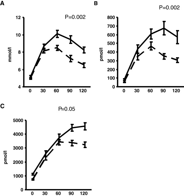
Figure 1.
(A-C): Glucose, insulin and C-Peptide levels during the oral glucose tolerance test. Data are presented as mean ± SE. Dashed lines represent participants with IHL ≤ 5.5%. Solid lines represent participants with IHL > 5.5%, i.e. NAFLD. P values are derived from linear regression modelling with the exposure IHL treated as a continuous variable and the outcome being the area under the curve for the relevant metabolite, adjusted for age, gender, alcohol consumption and visceral fat area.

