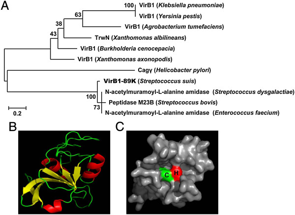
Figure 1.
Sequence analysis of VirB1-89K. (A) Phylogenetic analysis of VirB1-89K. Sequence alignment and phylogenetic analysis of VirB1-89K homologs were performed using MEGA 5.1 software. Values at nodes indicate bootstrap values for 500 replicates. (B) Analysis of the tertiary structure of the CHAP domain of VirB1-89K by using the online server SWISS-MODEL. (C) Visualization of the surface active site of the CHAP domain by using PyMOLviewer, showing the cysteine residue in green and histidine in red.

