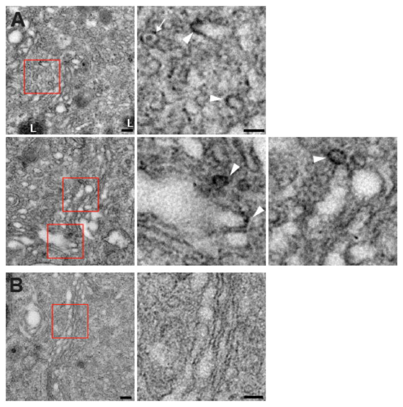
Figure 7. VPS41 Decorates Golgi-Derived Membrane Buds.

PC12 cells were transfected twice with VPS41 siRNA and once with cDNA encoding either siRNA-resistant miniSOG-VPS41 (A) or cytoplasmic miniSOG (B) alone as control. Three days after the second transfection, cells were fixed, oxidized by blue light in the presence of oxygen and DAB, and processed for EM.
(A) Electron micrographs show electron-dense DAB reaction product closely apposed to membrane buds associated with the ends of Golgi cisternae in cells transfected with miniSOG-VPS41. Red boxes indicate the regions magnified to the right. The white arrowheads indicate electron-dense deposits and the white arrow an unlabeled LDCV.
(B) A cell expressing the cytoplasmic miniSOG shows an unlabeled Golgi profile. Scale bars represent 200 nm. L, lysosome.
See also Figure S4.
