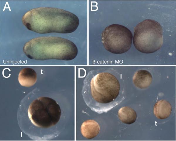
Fig. 3. Representative results of host transfers using X. laevis and X. tropicalis oocytes.
(A-B) Maternal knockdown of β-catenin. Oocytes injected with 10 ng of β-catenin MO were matured and fertilized via the host-transfer method, as in ref. (23). (A) Uninjected and (B) MO-injected embryos at stage 25 demonstrate the knockdown phenotype. Uninjected embryos develop normally while knockdown embryos are ventralized (100%, n=28). (C-D) Representative results of X. tropicalis oocytes fertilized following host-transfer into X. laevis females. (C) X. tropicalis embryo at the 4-cell stage (t) shown next to a host X. laevis embryo (l). (D) X. tropicalis embryos at the neurula stage (t) shown next to a host X. laevis embryo (l).

