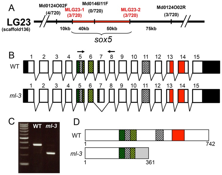
Figure 3. Mapping of the ml-3 locus.
(A) ml-3 locus was mapped to a 90 kb region (red bar) between MLG23-1 and MLG23-2 on LG23, predicted to contain only one gene, sox5. Typing markers are indicated above the map, with the number of recombinants per total 720 haploid genomes examined at each position. (B) Medaka sox5 comprises 15 exons (upper). Sequencing of cDNAs showed that in ml-3, exon 7 is skipped (lower). Boxes represent exons. Angled lines represent introns. The 5′ and 3′ untranslated regions are colored in black. Diagonal stripes and colored regions correspond to regions encoding the protein domains described in (D). Black arrows show positions of the primer set used for RT-PCR in C. (C) RT-PCR detects the skipping of exon 7 in sox5 mRNA of ml-3 mutant. (D) WT sox5 gene encodes 742 amino acid protein consisting of two coiled-coil domains (diagonal stripe) and HMG box (red box). The first coiled-coil domain contains a leucine-zipper (green box) and a glutamine-rich domain (Q box, light green box). In ml-3, loss of exon 7 causes a premature stop codon leading to a truncated protein. Gray box represents an altered frame. The resultant truncated protein would lack the second coiled-coil domain and HMG box (lower).

