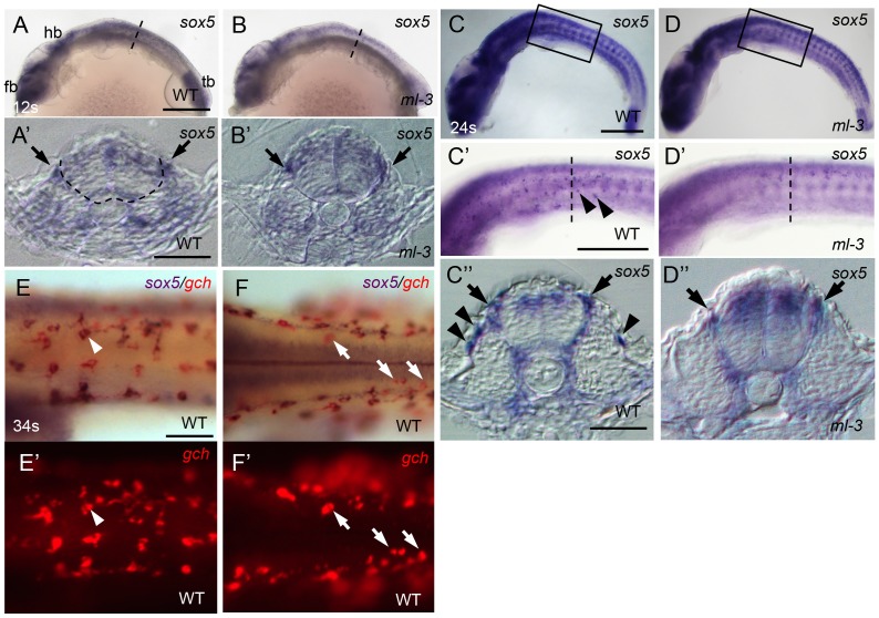
Figure 5. Expression pattern of medaka sox5 in WT and ml-3 mutant embryos.
(A, C, E, F) WT. (B, D) ml-3 mutant. (A′, B′, C″, D″) Transverse sections. (E, F) sox5 (blue) and gch (red). (A–E, E′) Lateral views. (F, F′) Dorsal views. (A) In WT embryos at 12 somite stage (12 s, 41 hpf), sox5 is expressed in premigratory NCCs (see also section in A′, black arrow) as well as in dorsal neural tube and CNS ranging from forebrain (fb) to hindbrain (hb), and in tailbud (tb). The boundary between neural tube and somite is indicated by dotted line. (B) In ml-3, the sox5 expression pattern is not markedly altered. In particular, when observed in section (B′), premigratory NCCs are positive for sox5. (C, C′, C″) At 24 somite stage (24 s, 58 hpf), sox5-expressing cells are found in dorsal neural tube, premigratory NCCs (arrows in C″) and migrating NCCs between neural tube and somite and lateral trunk surface (black arrowheads in C′, C″) pathways in WT. sox5-expressing cells scattered on lateral trunk surface are prominent in WT (C′, C″). (D, D′, D″) In ml-3, sox5 expressing cells are absent from lateral trunk surface (D′, D″), whereas sox5 expression remains in dorsal neural tube, premigratory NCCs (black arrows) and migrating NCCs between neural tube and somite (D″). Boxed portion in C, D are magnified in C′, D′, respectively. (A′, B′, C″, D″) Transverse histological section from embryos at the level as indicated by dotted line in A, B, C′ and D′. (E, E′) sox5-expressing cells on lateral trunk surfaces at 34 somite stage (34 s, 74 hpf) also express gch. White arrowhead represents an example of gch-positive sox5-expressing cell. (F, F′) On dorsal trunk surface, some sox5-negative gch-positive cells were detected (white arrows). Scale bars: (A, C) 200 µm; (C′) 100 µm; (A′, C″) 20 µm; (E) 50 µm.

