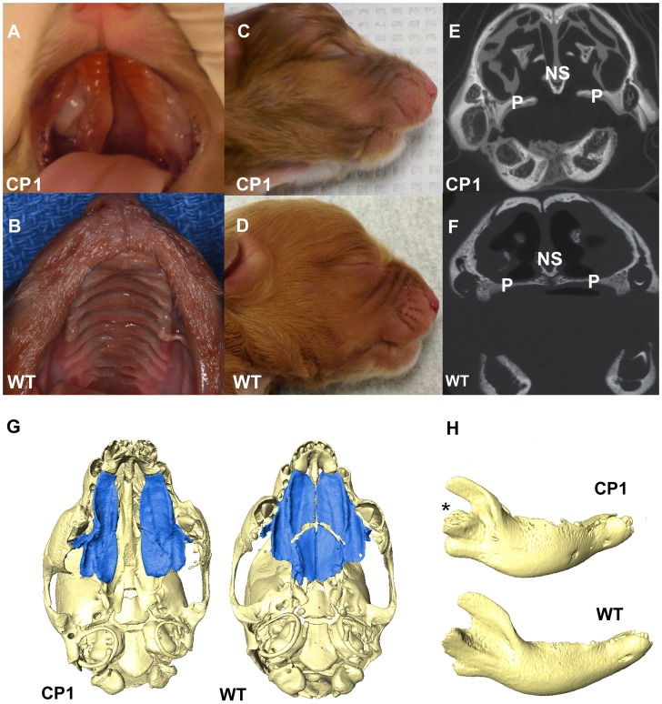
Figure 2. Phenotype of neonatal CP1 NSDTRs.
A. Neonatal CP1 NSDTR with an extensive cleft of the hard and soft palate. B. Neonatal NSDTR with a normal palate (WT). C. Lateral view of CP1 head exhibiting relative mandibular brachygnathia. D. Lateral view of WT head with a normal jaw relationship. E. Coronal CT image depicting the failure of the palatine processes and nasal septum to fuse in CP1 NSDTRs. F. Coronal CT image depicting midline fusion of palatal process and nasal septum in WT. P – Palatine process, NS – Nasal septum G. 3D reconstruction of microCT imaging of CP1 and WT skulls with mandibles removed. CP1 skull shows abnormally shaped palatine process and palatine bones. Bones colored blue are the palatine processes and palatine bones. WT skull shows anatomical location of normal palatine sutures and shape of palatine processes and palatine bones. H. 3D reconstruction of mandibles depicting abnormal angulation of the condylar process (*) in CP1 mandibles compared to WT mandibles.

