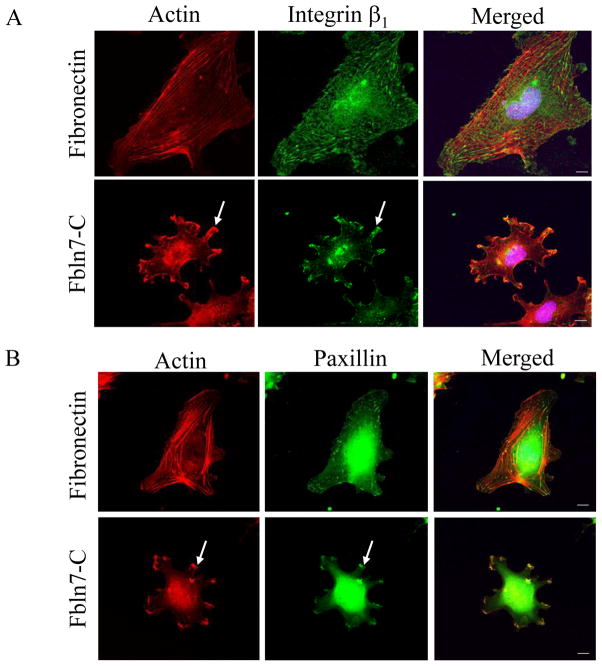
Fig. 2.
Accumulation of β1 integrin (A) and paxillin (B) in protrusions of HUVEC cells on Fbln7-C (20 μg/ml). Focal adhesions and actin stress fibers were well formed when the cells were plated on the fibronectin (5 μg/ml) substrate. On the Fbln7-C substrate, actin was accumulated at the tip of the protrusions, and β1 integrin and paxillin accumulated in clusters (arrows). Scale bar, 10 μm.

