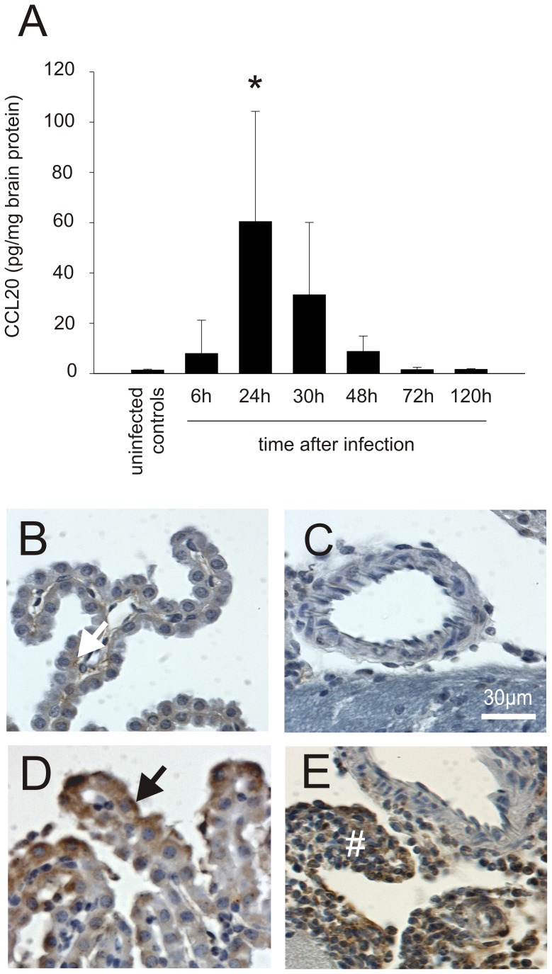
Figure 2. CCL20 is expressed mainly during acute pneumococcal meningitis.
(A) Increased CCL20 levels were found in mice brain homogenates during acute bacterial meningitis using ELISA. After initiation of antibiotic therapy (starting 24 h after infection), they decreased quickly to normal ((*) p<0.01 compared with uninfected controls). In uninfected control mice, (B, C) only a very subtle CCL20-positive staining was observed (white arrow). In contrast, in animals with pneumococcal meningitis, CCL20-positive staining was found in (D) epithel cells of the choroid plexus (black arrow) and (E) the subarachnoid inflammatory infiltrate (#). Number of animals: 6 h: n = 6, 24 h: n = 9, 30 h: n = 6, 48 h: n = 6, 72 h: n = 6, and 120 h: n = 7. Uninfected animals were used as controls (n = 11).

