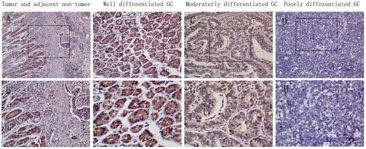
Figure 3. The in situ expression of UPK1A protein in gastric cancer specimens was assessed by immunohistochemistry.
(A) and (E), Immuno-staining of a GC tumor and the adjacent non-tumorous area. (B) and (F), Strong UPK1A staining was observed in well-differentiated GC. (C) and (G), Weak UPK1A staining in moderately differentiated gastric cancer. (D) and (H), Negative UPK1A staining in poorly differentiated GC. (A–D with 200× magnification; E–H with 400×magnification).

