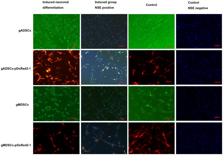
Figure 3. Neuronal differentiation of gADSCs, gADSCs–pDsRed2-1, gMDSCs and gMDSCs–pDsRed2-1.
gADSCs, gADSCs–pDsRed2-1, gMDSCs and gMDSCs–pDsRed2-1 were altered morphologically by incubation in induction medium containing β-mercaptoethanol. The cytoplasm began to shrink, and after 2 h the cell bodies became conical, triangular or round in shape with multiple protrusions resembling axons, the ends of which were primary and secondary bifurcated dendrites. Immunohistochemistry showed that the cells were NSE (green) positive after 3 h. Uninduced control cells were morphologically unaltered and did not express NSE. gADSCs, goat adipose-derived mesenchymal stem cells; gMDSCs, goat muscle-derived satellite cells; NSE, neuron-specific enolase.

