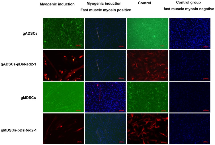
Figure 4. Myogenic induction of gADSCs, gADSCs–pDsRed2-1, gMDSCs and gMDSCs–pDsRed2-1.
gMDSCs and gMDSCs–pDsRed2-1 began to fuse after 2 d and myotube cells increased markedly. gADSCs and gADSCs–pDsRed2-1 began to integrate 5 d after myogenic induction, leading to short myotube cell formation. DAPI staining revealed the presence of multiple nuclei in the same myotube, and immunohistochemical staining demonstrated that the cells were fast muscle myosin (green) positive; control (uninduced) cell were fast muscle myosin negative. gADSCs, goat adipose-derived mesenchymal stem cells; gMDSCs, goat muscle-derived satellite cells.

