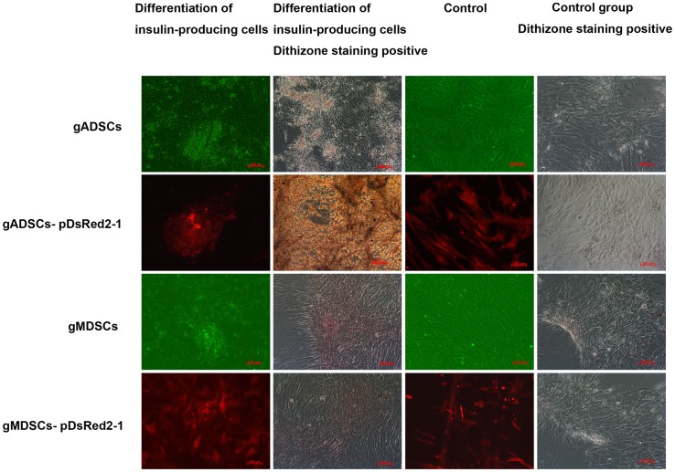
Figure 5. Differentiation and confirmation of insulin-producing cells.
gADSCs, gADSCs–pDsRed2-1, gMDSCs and gMDSCs–pDsRed2-1 exhibited intensive cell growth. There were irregular cell clusters in the control group, and the cells were divergent and fusiform. Cell masses were scarlet in the induced group after dithizone staining but remained unstained in the control group. Insulin was detected in fusiform cells in the induced group, but the control group lacked insulin. gADSCs, goat adipose-derived mesenchymal stem cells; gMDSCs, goat muscle-derived satellite cells.

