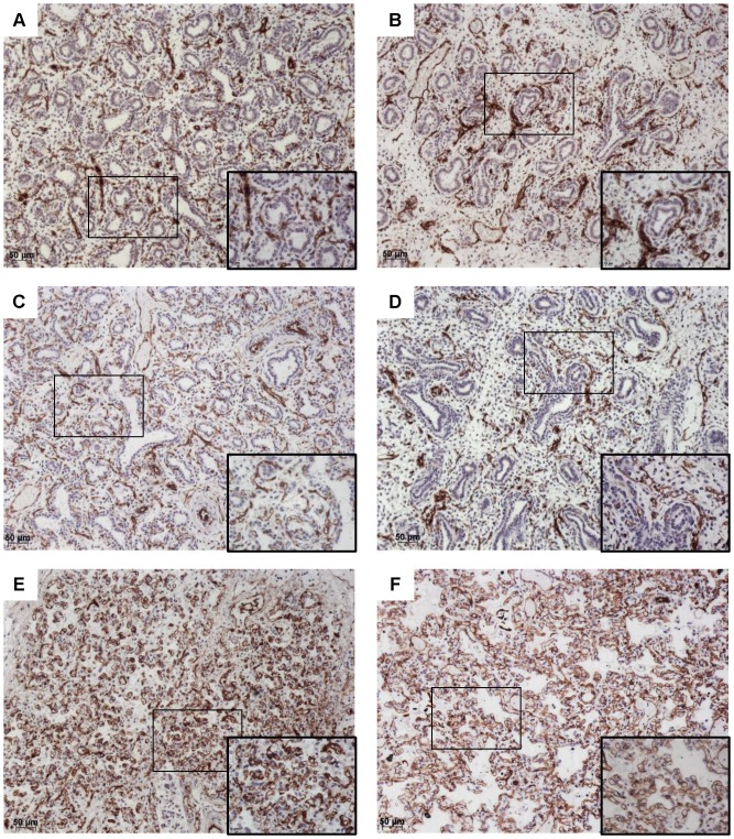
Figure 3. CD31 immunohistochemistry.
Original magnification×10, and ×40 magnification of the area identified by a rectangle. CD31 (brown) and counterstaining with hematoxylin. Fetuses with PH (A, C, E) were compared with fetuses of a similar gestational age without PH (B, D, F). A: Fetus with right ventricular hypoplasia and a septal defect, 18 weeks, LW/BW = 0.010; B: Fetus with pulmonary atresia and a septal defect, 16 weeks, LW/BW = 0.024; C: Fetus with tetralogy of Fallot, 22 weeks, LW/BW = 0.005; D: Fetus with an atrioventricular septal defect, 17 weeks, LW/BW = 0.027; E: Fetus with pulmonary atresia and tricuspid atresia, 36 weeks, LW/BW = 0.009; F: Fetus with tetralogy of Fallot, 33 weeks, LW/BW = 0.029.

