
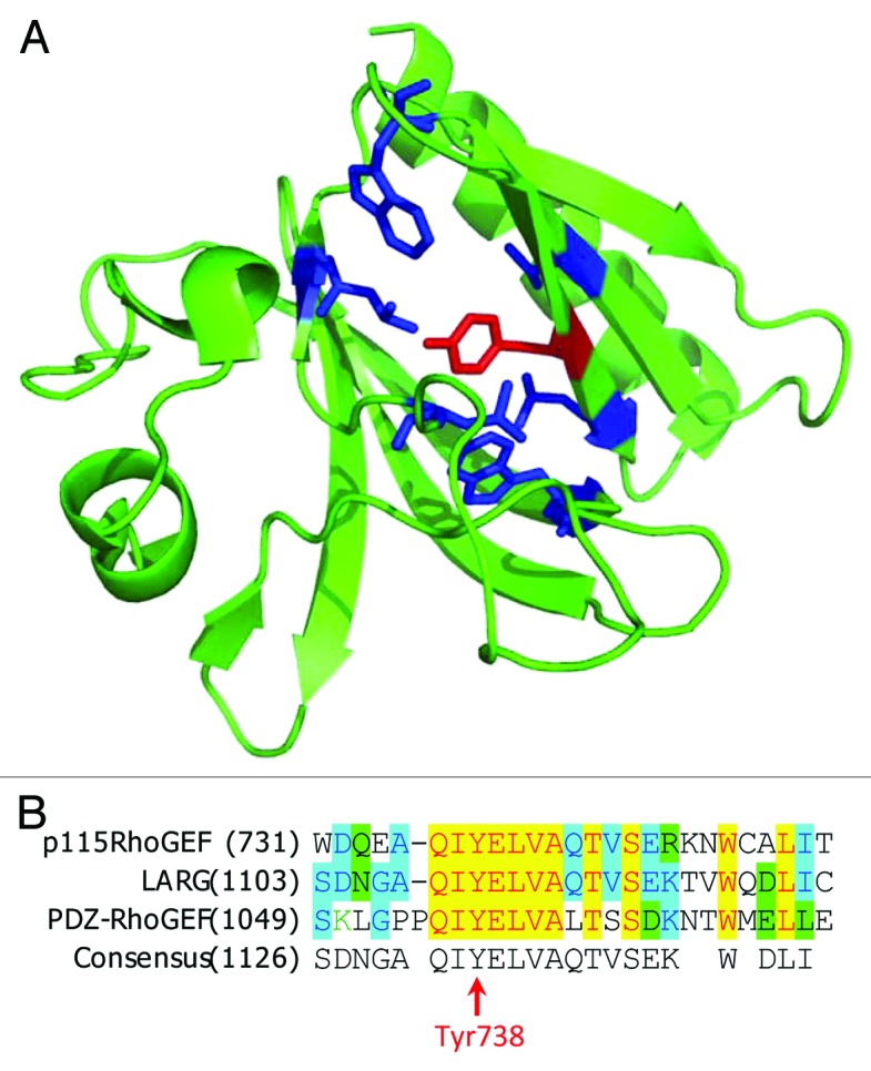
Figure 3. Phosphorylation of PH domain of p115RhoGEF by JAK2. (A) Structure of p115RhoGEF’s PH domain (PDB identifier: 3ODO). TheTyr738 residue is depicted in red and the surrounding hydrophobic residues are depicted in blue. (B) Alignment of amino acid sequences for p115RhoGEF, LARG, and PDZ-RhoGEF.