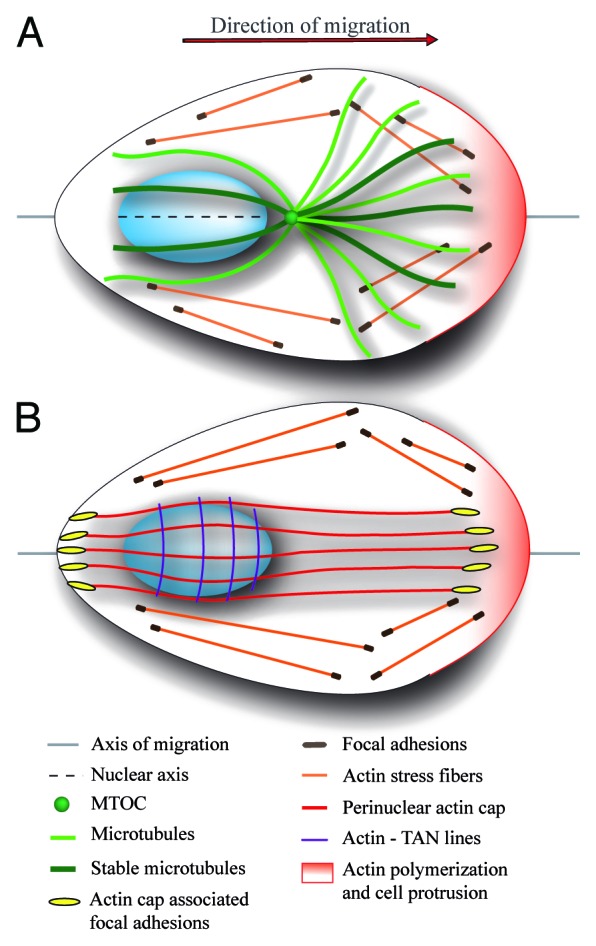
Figure 1. Schematic representation of an adherent migrating cell with front-rear polarity. Polarized cell displays conical shape with actin polymerization induced at the leading edge (pink) and limited at the cell rear. Cells are attached to the substrate through cell–matrix adhesion, such as focal contacts and adhesions, which connects extracellular matrix to cellular cytoskeleton. (A) In polarized cells, the oval-shaped nucleus localizes to the cell rear, MTOC in front of the nucleus close to the cell center, and microtubules are preferentially oriented toward the leading edge and they are stabilized at this location. The relative position of the nucleus and MTOC is an important marker of cell migration polarity defining the axis of migration. In addition, the longer nuclear axis is aligned with the axis of migration. (B) Specific types of actin filaments anchored to the dorsal side of the nucleus contribute to the establishment of asymmetric profile of the migrating cell. TAN lines are arranged perpendicular to axis of migration and drive the rearward movement of the nucleus during cell polarization. Perinuclear actin cap fibers span the nucleus and link the nucleus to subset of focal adhesions at cell periphery. Actin cap filaments are aligned with the axis of migration and longer nuclear axis and they probably stabilize nuclear orientation. It is not clear whether perinuclear actin cap and TAN lines co-exist in the same cell.
