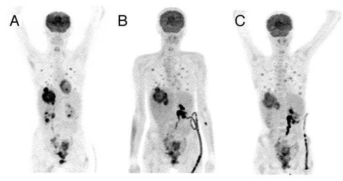
Figure 1. Functional imaging at different treatment time points. [F-18]2-deoxy-2-fluoro-d-glucose positron emission tomography (F-18-FDG PET/CT) maximum intensity projection images (antero-posterior view) demonstrating the FDG uptake during the treatment course of the patient. Upon inclusion (A) high accumulation of FDG was shown within the target lesion in the liver. Quantitative evaluation of FDG uptake of the first follow-up scan after three weeks of lenalidomide monotherapy (B) revealed a reduction of the maximum standard uptake value (SUVmax) within the target lesion by 45%, from initially 12.1 to 6.7. Restaging after three weeks of combined lenalidomide and cetuximab treatment showed a slight increase of the SUVmax to 7.6.
