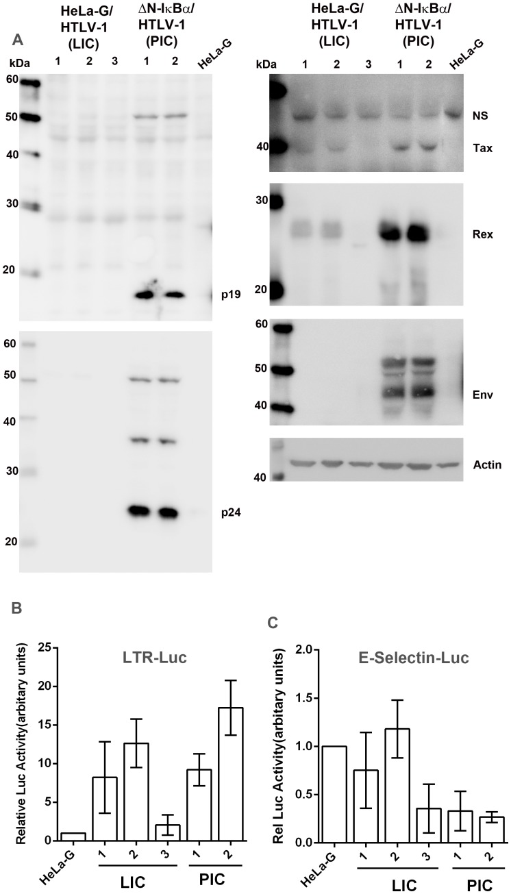
Figure 2. Characterizations of HeLa-G-derived cell lines productively (PIC, ΔN-IκBα/HTLV-1) or latently infected (LIC, HeLa-G/HTLV-1) by HTLV-1.
(A) Immunoblot analyses of whole-cell lysates of HeLa-G/HTLV-1 clones 1, 2, and 3 (LIC); HeLa-G/ΔN-IκBα/HTLV-1 (ΔN-IκBα/HTLV-1, PIC) clones 1 and 2; and HeLa-G control. Antibodies used include p19, p24, Tax, Rex, Env, and β-actin (Actin). (B & C) LTR-Luc and E-selectin- Luc reporter activities in HTLV-1-infected cell lines analyzed in (A). Each reporter plasmid and the control Renilla-luciferase plasmid, pRL-TK, were transfected into HeLa-G cells, 3 LIC lines and 2 PIC lines as described in Materials and Methods . Relative luciferase activity after normalization is plotted.

