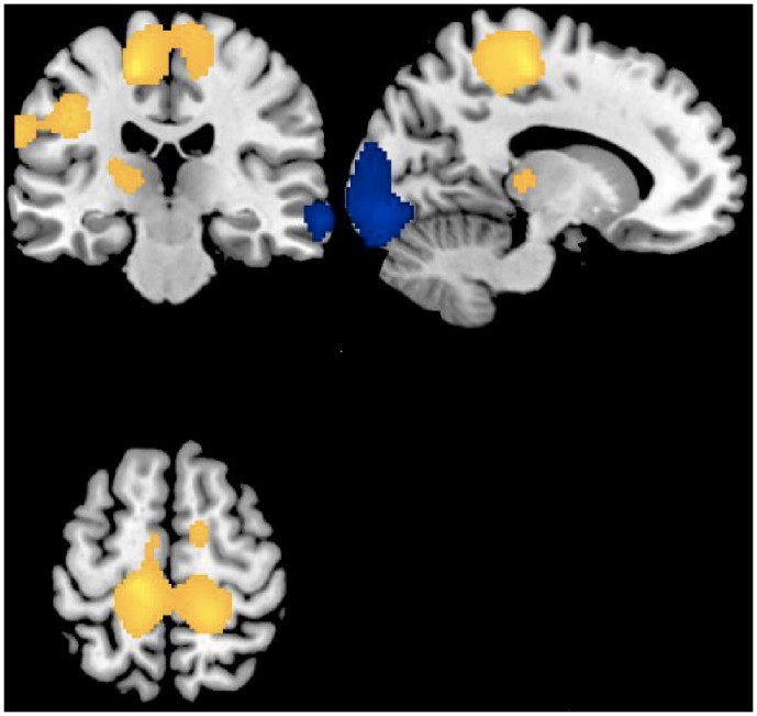
Figure 1. Regional cerebral increases (yellow blobs) and decreases (blue blobs) in Kleine-Levin patients (n = 4) during symptomatic episodes as compared to baseline asymptomatic period, superimposed on a template T1-weighted MRI.
Activations are displayed at p<.05, uncorrected, for clusters >50 voxels.

