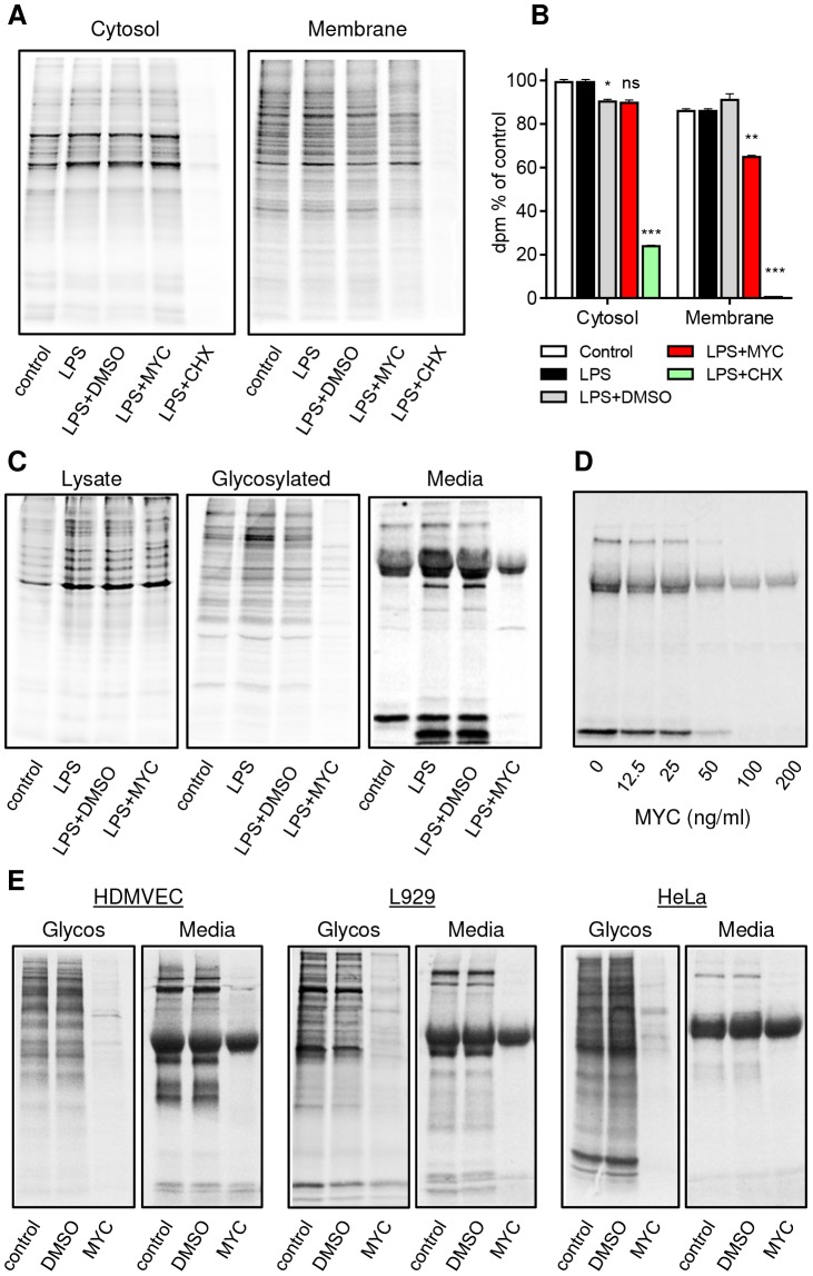
Figure 6. Mycolactone specifically targets membrane and secreted proteins.
Cells in Met/Cys free medium were incubated +/−125 ng/ml mycolactone (MYC), 10 µg/ml cycloheximide (CHX) or 0.0125% DMSO for 1 hr and stimulated with LPS for 1 hr before replacement with fresh medium containing Tran35slabel for 2 hrs. Data representative of 3 independent experiments (A–D). A. Cytosolic and digitonin-resistant membrane fractions from treated RAW264.7 cells (105 cell equivalents/lane). B. Quantification of 35S incorporation in (A) by scintillation counting (mean±SEM, n = 3). *, P<0.05; **, P<0.01; ***, P<0.001. C. Total cell lysates, Concanavalin A (ConA) agarose precipitated proteins (Glycosylated) and supernatants (Media) from RAW264.7 cells. D. Dose dependence of the suppression of supernatant protein production in RAW264.7 cells. E. ConA precipitated (Glycos) and supernatant proteins (Media) from 35S labelled Human microvascular dermal epithelial cells (HDMVEC), L929 fibroblasts and Hela cells. In each case total cell labelling was comparable between samples (not shown).

