
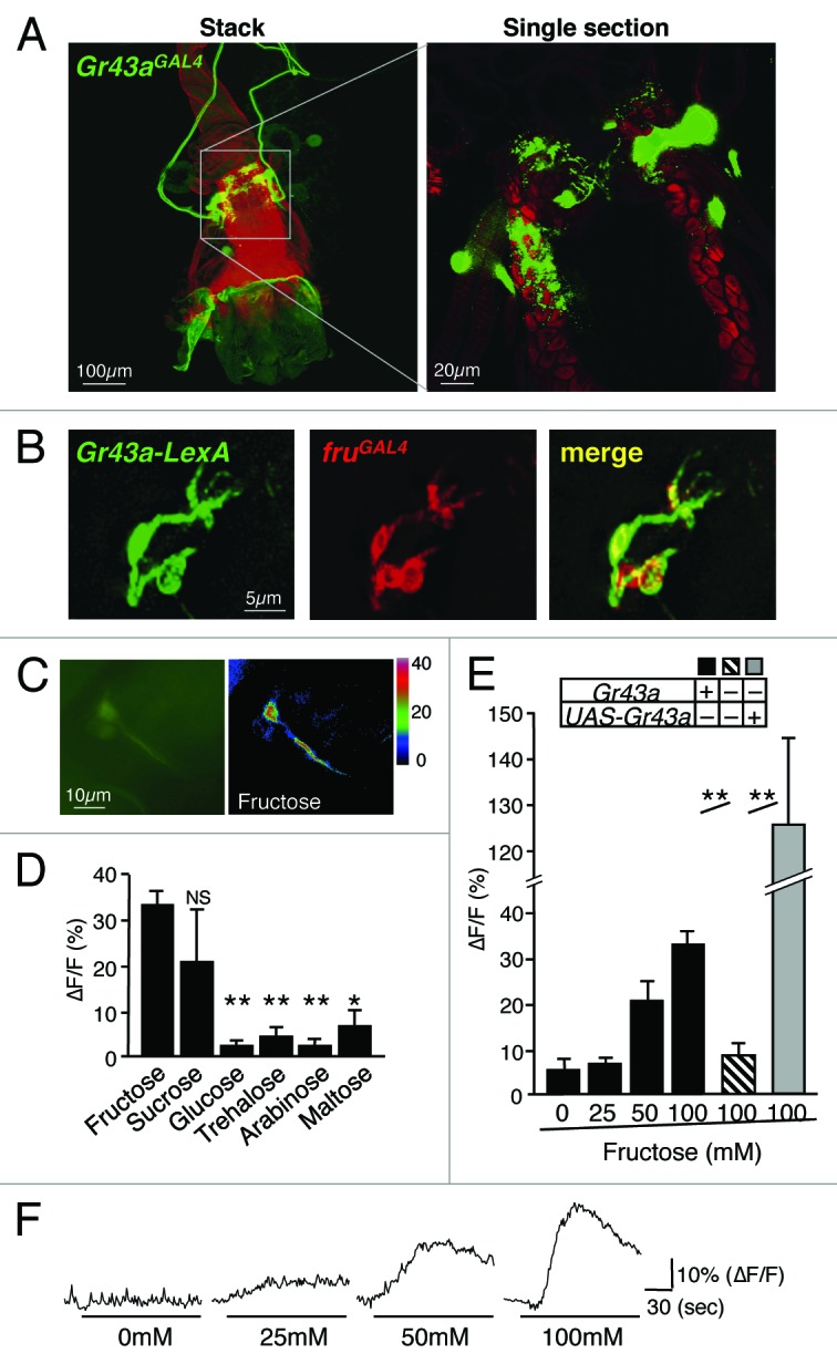
Figure 3.Gr43a expressing neurons in the uterus. (A) Gr43aGAL4 drives UAS-mCD8GFP expression in 4–6 neurons. Cell bodies are located at the middle of the uterus, sending the axons to the abdominal ganglion. The dendrites innervate the lumen of the uterus, clearly shown by the single section view. (B) Gr43a-LexA and SPR are co-expressed in the same neurons. Gr43a-LexA drives lexAop-rCD2GFP, fruGAL4 drives UAS-mCD8RFP. Note that fruGAL4 expression serves as a proxy for SPR, which are co-expressed in the uterus.35,36 (C) Uterus neurons expressing G-CaMP3.0 under control of Gr43aGAL4. ∆F pseudocolor fluorescence image was taken 85 s after application of 100mM fructose (right). (D) Gr43aGAL4 neurons specifically respond to fructose. Max ∆F/F within 135 s of application is shown. All sugars are 100mM. Flies contained 2 genomic copies of Gr43a. **P < 0.0001; ANOVA. Error bars represent standard error. 5 ≤ n ≤ 8. (E) Response of Gr43aGAL4 neurons to fructose is Gr43a dependent. **P < 0.0001; ANOVA. Error bars represent standard error. 5 ≤ n ≤ 9. (F) Time-course of G-CaMP3.0 fluorescence changes in Gr43aGAL4 neurons stimulated with different concentrations of fructose. Note that ∆F/F in Gr43aGal4/Gr43aGAL4;UAS-Gr43a rescue flies is higher than in Gr43a+ controls, likely due to higher expression levels of Gr43a.