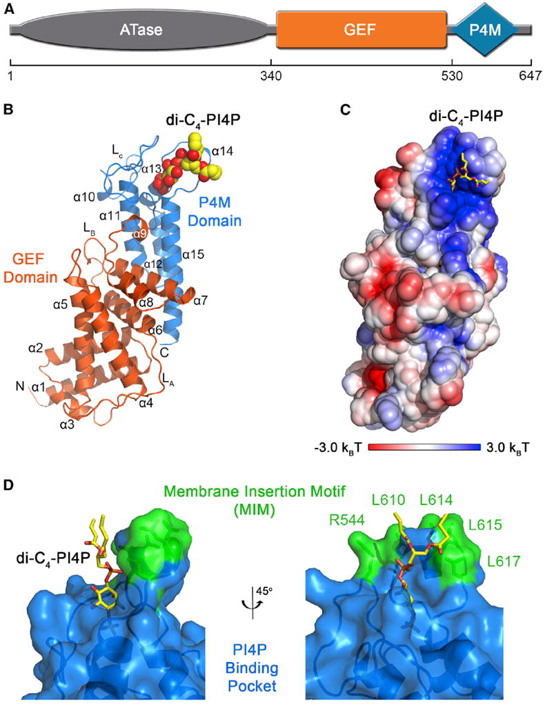
Figure 2. Crystal Structure of DrrA330–647 in Complex with Dibutyl PI(4)P.

(A) Domain architecture of DrrA.
(B) Overall view with the GEF and P4M domains colored as indicated and dibutyl PI(4)P depicted as spheres. Secondary structural elements are numbered starting with the first helix of the GEF domain. di-C4-PI4P, dibutyl PI(4)P.
(C) Surface representation of the P4M domain colored according to electrostatic potential calculated with APBS (Baker et al., 2001). Dibutyl PI(4)P is shown as sticks.
(D) View of the PI(4)P binding pocket with DrrA rendered as ribbons with a semitransparent surface. Dibutyl PI(4)P and side chains in the putative membrane insertion motif are shown as sticks.
