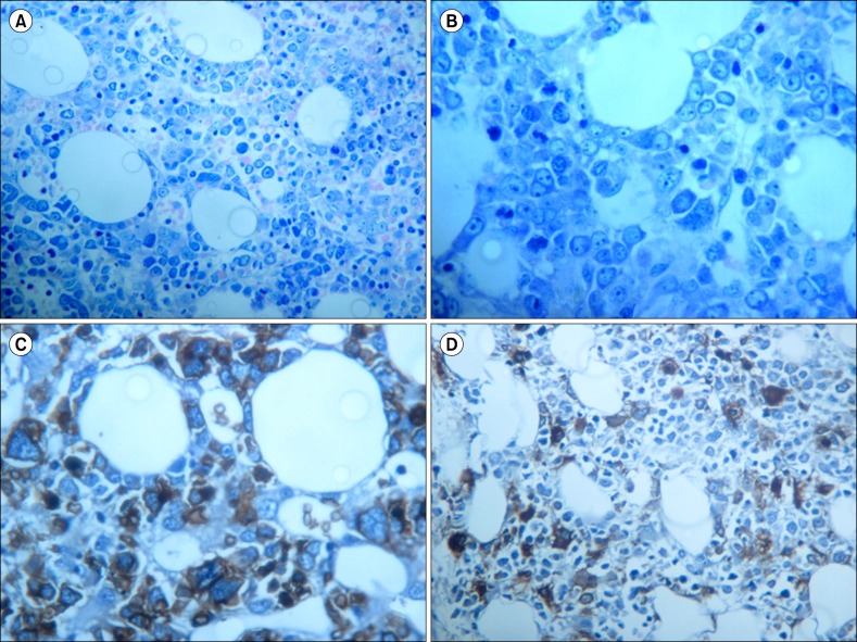
Fig. 2.
Bone marrow trephine biopsy at diagnosis of pure erythroid leukemia. (A) Medium magnification (×200) shows a predominant bone marrow population composed of atypical immature erythroid precursors (more than 90% with nucleated cells). (B) High magnification (×400) revealed numerous atypical erythroid precursors and dyserythropoiesis. (C) Glycophorin-A immunostain expressed by the atypical erythroid neoplastic precursors. (D) Linker for activated T-cells immunostain shows dysmegakaryocytopoiesis with a micromegakaryocytes and a population of immature megakaryoblasts.

