
Figure 1.
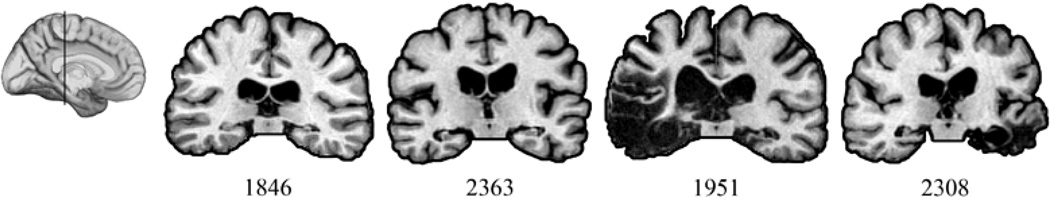
Magnetic resonance scans of hippocampal patients. Images are coronal slices through the midportion of the hippocampus from T1-weighed scans. Volume changes can be noted in the region of the hippocampus bilaterally.
Official websites use .gov
A
.gov website belongs to an official
government organization in the United States.
Secure .gov websites use HTTPS
A lock (
) or https:// means you've safely
connected to the .gov website. Share sensitive
information only on official, secure websites.
Magnetic resonance scans of hippocampal patients. Images are coronal slices through the midportion of the hippocampus from T1-weighed scans. Volume changes can be noted in the region of the hippocampus bilaterally.