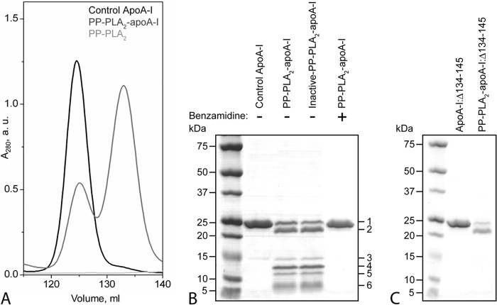
FIGURE 4.
Characterization of lipid-free apoA-I and apoA-I:Δ134–145 after reaction with PP-PLA2. Purified plasma apoA-I was incubated with PP-PLA2 at an enzyme/substrate ratio of 1:200 at 25 °C for 1 h. Control samples represent apoA-I samples incubated under the same conditions in the absence of PP-PLA2. A, SEC chromatograms of control apoA-I, PP-PLA2-apoA-I, and PP-PLA2 only. For SEC analysis, samples were loaded at 0.5 mg/ml (in total protein concentration) on a preparative grade Superdex 200 XK 16/100 column and eluted with 10 mm PBS, pH 7.5, at a flow rate of 0.5 ml/min. B, SDS-PAGE of control apoA-I, PP-PLA2-apoA-I, and inactive PP-PLA2-apoA-I, in which PP-PLA2 was preincubated with EDTA before reaction with apoA-I. 5 μg of protein was loaded per lane. Bands 1–6 represent proteolyzed fragments of apoA-I that were excised from the gel and analyzed by MALDI-TOF MS. C, SDS-PAGE of apoA-I:Δ134–145 and PP-PLA2-apoA-I:Δ134–145. Molecular weight standards are Precision Plus Protein Dual Xtra standards (Bio-Rad). a.u., absorbance units.

