FIGURE 2.
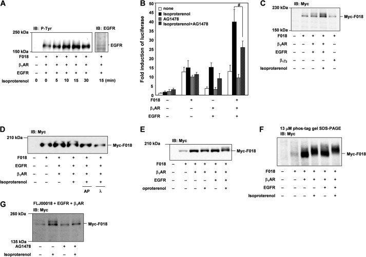
FLJ00018 is activated by β1-AR-mediated transactivation of EGF receptors. A, NIH3T3 cells were co-transfected with expression vectors for EGFR, β1-AR, and FLJ00018 (F018) as indicated. Transfected cells were stimulated with isoproterenol (20 μm) for 0–30 min. Equal amounts of protein were resolved by 7.5% SDS-PAGE. Immunoblotting (IB) was performed with antibodies against phosphotyrosine (P-Tyr) or EGFR. B, NIH3T3 cells were co-transfected with pSRE.L-luciferase, pRL-SV40 plasmid DNAs, and expression vectors for FLJ00018, β1-AR, and EGFR as indicated. Transfected cells were stimulated with 20 μm isoproterenol for 5.5 h before treatment with 10 μm AG1478 for 1 h. Luciferase activity was determined by a dual-luciferase reporter assay. Luciferase activity obtained with mock cells was taken as 1.0, and relative activities are shown. Values are the means ± S.D. from at least three experiments. #, p < 0.05. C, NIH3T3 cells were co-transfected with expression vectors for EGFR, β1-AR, Myc-tagged FLJ00018, and Gβ1γ2 as indicated. Transfected cells were stimulated with isoproterenol (20 μm) for 15 min. Equal amounts of protein were resolved by 7.5% SDS-PAGE. To detect Myc-tagged FLJ00018, immunoblotting was performed with antibodies against Myc (Myc). D, NIH3T3 cells were co-transfected with expression vectors for EGFR, β1-AR, and Myc-tagged FLJ00018 as indicated. Transfected cells were stimulated with isoproterenol (20 μm) for 15 min. After lysis, the cell lysates were incubated with alkaline phosphatase (AP; 10 units) and λ-phosphatase (λ; 400 units) for 2 h at 30 °C. The reaction was terminated by the addition of SDS sample buffer. Equal amounts of the reaction mixture were resolved by 7.5% SDS-polyacrylamide gel electrophoresis. To detect Myc-tagged FLJ00018, immunoblotting was performed with antibodies against Myc (Myc). E and G, HEK293 cells (E) or NIH3T3 cells (G) were co-transfected with expression vectors for EGFR, β1-AR, and Myc-tagged FLJ00018. Transfected cells were treated with 10 μm AG1478 for 30 min (G) before stimulation with 20 μm isoproterenol for 15 min. Equal amounts of protein were resolved by 7.5% SDS-PAGE (E and G) or 3% SDS-polyacrylamide gel containing 13 μm Phos-tag, 26 μm MnCl2 and 1.5% agarose (F). Immunoblotting was performed with anti-Myc.

