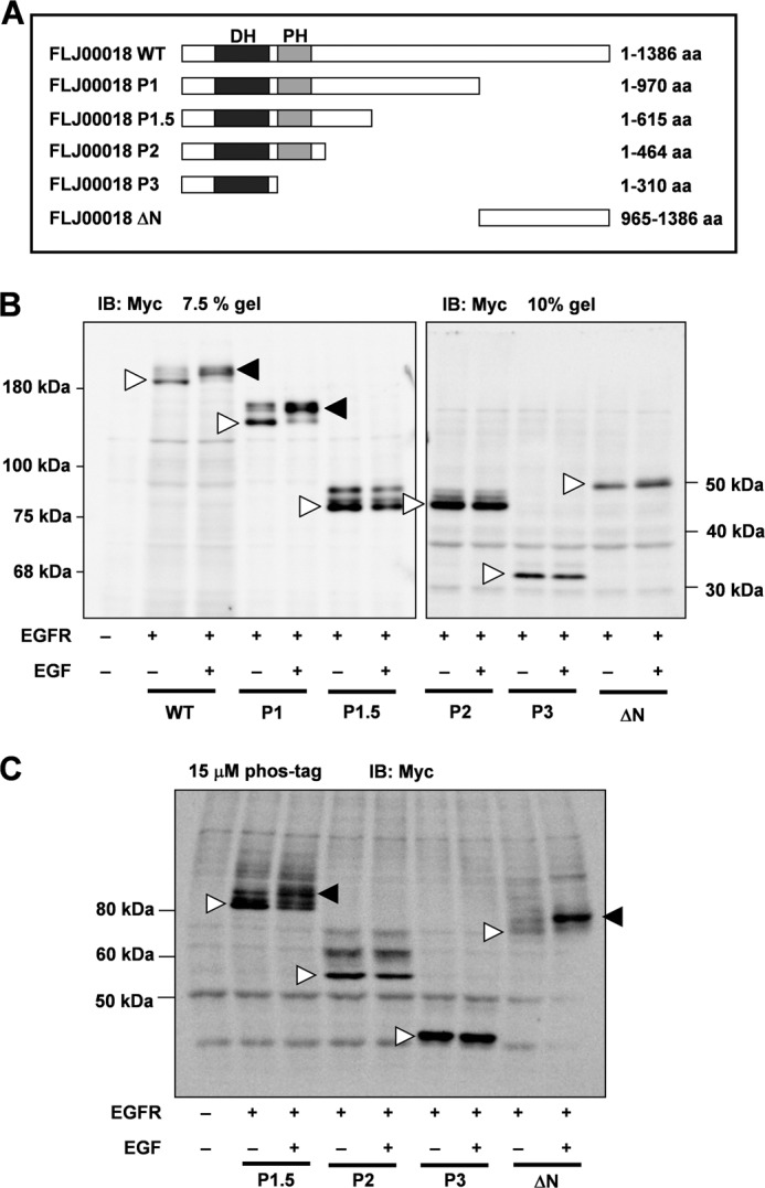
FIGURE 6.

Identification of the structure involved in FLJ00018 phosphorylation by EGFR signaling. A, structure of FLJ00018 mutants. The WT, P1, P1.5, P2, P3, and ΔN constructs code for amino acid (aa) residues 1–1386, 1–970, 1–615, 1–464, 1–310, and 965–1386 of FLJ00018, respectively. B and C, NIH3T3 cells were co-transfected with expression vectors for EGFR and Myc-tagged FLJ00018 mutants as indicated. Transfected cells were stimulated with 20 ng/ml EGF for 15 min. Equal amounts of proteins were resolved by 7.5% or 10% SDS-PAGE (B) or 6% SDS-polyacrylamide gel containing 15 μm Phos-tag and 30 μm MnCl2 (C). Immunoblotting (IB) was performed with antibodies against Myc. Closed triangles, shifted bands; Open triangles, basal levels of proteins.
