
FIGURE 8.
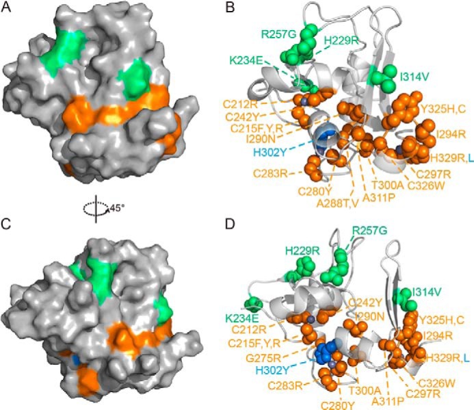
Location and structural effects of previously identified PHF6-ePHD2 domain mutations. A–D, surface (A and C) and sphere (B and D) representations of PHF6-ePHD2 domain mutations that found in BFLS (green), in acute myeloid leukemia (blue), and in T-ALL (orange). Zinc ions are in gray.