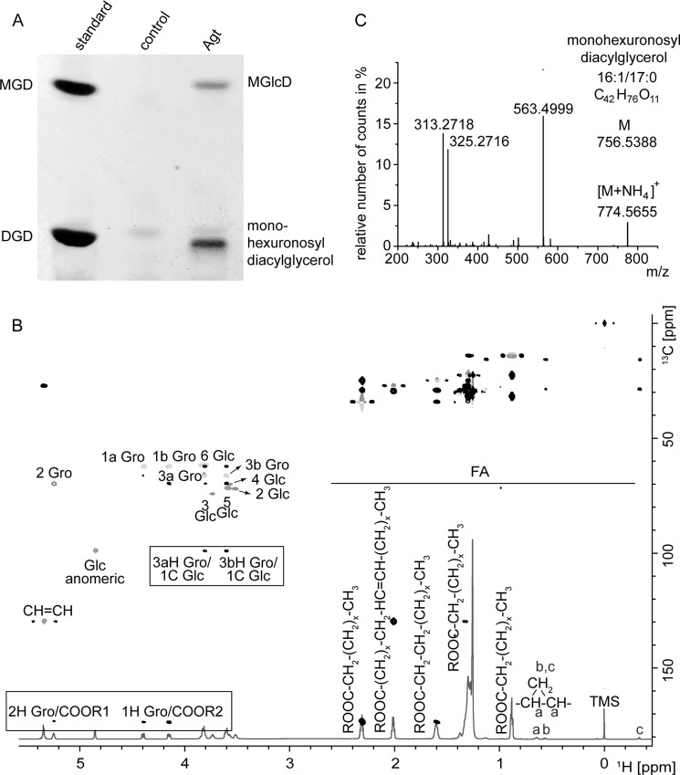
FIGURE 1.
Accumulation of two new glycolipids in E. coli expressing Agt from Agrobacterium. A, TLC of lipid extracts from E. coli BL21 (DE3) expressing Agt or the empty vector as control. The new glycolipids were identified as MGlcD and monohexuronosyl diacylglycerol. B, overlay of 1H,HSQCdept and HMBC spectra of MGlcD. Spectra were recorded in CDCl3 at 27 °C utilizing the Bruker DRX Avance 700 MHz spectrometer. Important intra-residual scalar correlations are marked in the box. R1 and R2 indicate the following: 14:0; 16:1; 16:0; 17:0c (ω9,10); 18:1; 19:0c (ω9,10). TMS, tetramethylsilane; FA, fatty acids. C, Q-TOF MS/MS spectrum of monohexuronosyl diacylglycerol. Parental ions were selected as ammonium adducts and fragmented. The figure shows the spectrum of one main species with m/z 774.5727. The fragment with m/z 563.4999 represents DAG-16:0/17:0c (as protonated form with loss of H2O). The neutral loss of 211.0660 (m/z 774.5655 minus 563.4999) is derived from hexuronic acid (as ammonium adduct). The ions m/z 313.2718 and 325.2716 represent monoacylglycerol-16:0 and monoacylglycerol-17:0c, respectively, each as protonated form with loss of H2O.

