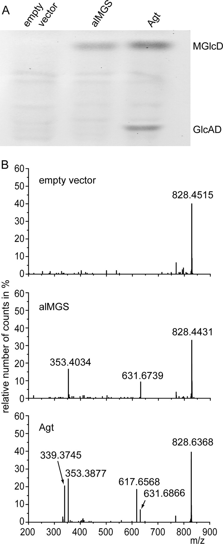
FIGURE 4.

Accumulation of MGlcD and GlcAD in Agrobacterium double knock-out mutant Δagt Δpgt transformed with the empty vector, alMGS from Acholeplasma or Agt from Agrobacterium. A, TLC of lipid extracts from the three lines with expression of alMGS leading to the synthesis of MGlcD and Agt to the synthesis of both MGlcD and GlcAD. B, Q-TOF MS/MS spectra of MGlcD and GlcAD. Parental ions with a calculated m/z 828.6196 (with a detection window m/z 1.2) were selected in the positive mode. Because of the similarity of their m/z values, ions representing ammonium adducts of both GlcAD-18:1/19:0c (calculated m/z 828.6196) and MGlcD-19:0c/19:0c (calculated m/z 828.6560) were selected together for fragmentation in this experiment. The fragments with m/z 617.6 and 631.6 represent DAG-18:1/19:0c and DAG-19:0c/19:0c (as protonated form with loss of OH), respectively. The neutral loss of 197.0 (m/z 828.6 minus 631.6) is derived from glucose (as ammonium adduct); the neutral loss of 211.0 (m/z 828.6 minus 617.6) is derived from glucuronic acid (as ammonium adduct). Therefore, DAG-18:1/19:0c is derived from GlcAD, whereas DAG-19:0c/19:0c is derived from MGlcD. The peaks at m/z 353.3877/353.4034 or 339.3745 represent protonated monoacylglycerol (MAG) species containing a 19:0c or 18:1 fatty acid, respectively, with loss of H2O. The molecular species of MGlcD and GlcAD shown in the spectra represent one example each of different molecular species of the two glycolipids detected in Agrobacterium with all showing the same result that GlcAD is only present in cells expressing Agt and absent in cells expressing alMGS from Acholeplasma.
