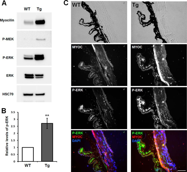
FIGURE 10.
Enhanced ERK activation in the trabecular meshwork of myocilin-expressing transgenic mice. A, Western blot analysis of the indicated proteins in the eye angle tissues. Lysates from the dissected angle tissues of 8-month-old wild-type or transgenic mice were immunoblotted with anti-myocilin (1:2,000 dilution), anti-phospho-MEK (P-MEK; 1:500 dilution), anti-phospho-ERK (P-ERK; 1:500 dilution), anti-total ERK (1:1,000 dilution), and anti-HSC70 (1:2,000 dilution) antibodies. B, quantification of the results obtained with four pairs of mice for phospho-ERK1/2. Error bars, S.D. C, eye sections of 8-month-old wild-type and transgenic mice were stained with anti-phospho-ERK (1:100 dilution), anti-myocilin (1:100 dilution) antibody, and DAPI. Top row, bright field images. TM, trabecular meshwork; CB, ciliary body. Scale bar, 50 μm.

