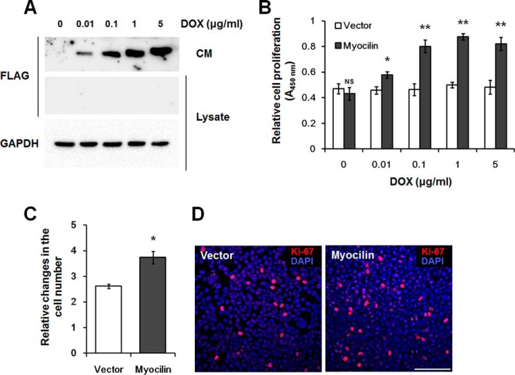
FIGURE 3.
Myocilin stimulates division of Tet-On HEK293 cells. A, Western blot analysis of myocilin in CM and cell lysates. Myocilin expression was induced by the addition of indicated concentration of DOX for 48 h. Anti-FLAG antibodies were used for detection of myocilin. Staining of the same blot with antibodies against GAPDH was used for normalization of loading. B, WST-1 proliferation assay. Increasing DOX concentrations were added to control (Vector) and myocilin-expressing Tet-On HEK293 cells for 48 h. The WST-1 reagent was added to each well. After 1 h, the absorbance at 450 nm was measured to evaluate cell proliferation. C, relative changes in the cell number after incubation of control and myocilin-expressing cells in the presence of 1 μg/ml DOX for 48 h. The equal numbers of cells (1 × 10⁵ cells/well) were plated into 6-well plates, and the increased numbers of cells were calculated as -fold changes relative to initial plating numbers. Error bars, S.D. of triplicate cultures. D, immunostaining of control and myocilin-expressing Tet-On HEK 293 cells with Ki-67 antibodies. Cells were plated in triplicate cultures and grown for 48 h in the presence of 1 μg/ml DOX. Nuclei were stained with DAPI. Scale bar, 100 μm (NS, non-significant; *, p < 0.05; **, p < 0.01).

