FIGURE 7.
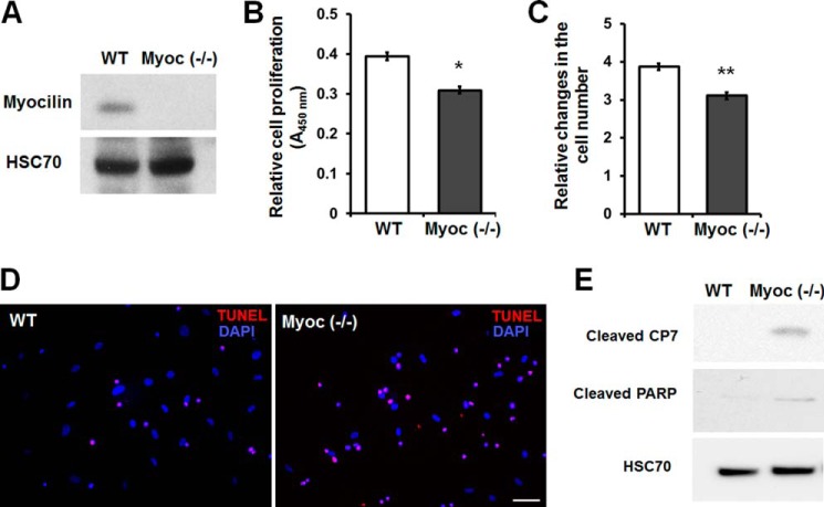
Myoc-null MSCs exhibit reduced cell proliferation and increased sensitivity to serum deprivation-induced apoptosis. A, Western blot analysis of MSCs isolated from wild-type or Myoc-null mice. Cell lysates were immunoblotted with anti-myocilin and anti-HSC70 antibodies. HSC70 was used for normalization of loading. B, WST-1 proliferation assay. Wild-type and Myoc-null MSCs were incubated for 48 h. Error bars, S.D. of triplicate cultures. C, relative changes in the cell number after incubation of wild-type and Myoc-null MSCs for 48 h. Equal numbers of cells (7 × 10⁴ cells/well) were plated into 6-well plates, and changes in the number of cells were calculated as -fold changes relative to the initial plating numbers. Error bars, S.D. of triplicate cultures. D, apoptosis in wild-type and Myoc-null MSC cultures after incubation in serum-free medium for 120 h. Apoptotic cells (red fluorescence) were identified by TUNEL assay. Scale bar, 100 μm. E, Western blot analysis of cell lysates using antibodies against cleaved CP7, cleaved poly(ADP-ribose) polymerase, and HSC70. Cells were incubated as in D. Similar results were obtained in three independent experiments. Representative blots are shown. *, p < 0.05; **, p < 0.01.

