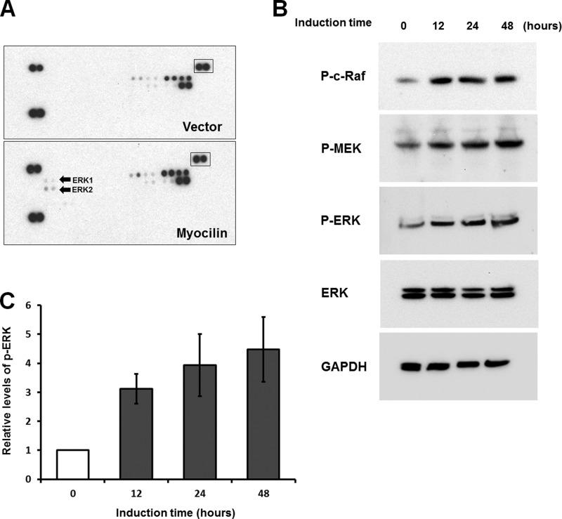
FIGURE 8.
Analysis of MAPK phosphorylation. A, control and myocilin-expressing Tet-On HEK293 cells were treated with 1 μg/ml DOX for 48 h. Lysates of control (top) and myocilin-expressing cells (bottom) were added to human phospho-MAPK antibody arrays. Arrays were treated as described under “Experimental Procedures.” Arrows, spots corresponding to phosphorylated ERK1 and ERK2 on the blots. Boxed spots were used for normalization. B, Western blot analysis of myocilin-expressing Tet-On HEK 293 cells. Cells were treated with 1 μg/ml DOX for the indicated periods of time. Cell lysates were immunoblotted with anti-phospho-c-Raf (P-c-Raf; 1:500 dilution), anti-phospho-MEK (P-MEK; 1:500 dilution), anti-phospho-ERK (P-ERK; 1:500 dilution), anti-total ERK (1:1,000 dilution), and anti-GAPDH (1:2,000 dilution) antibodies. C, quantification of the results of three independent experiments as in B for phospho-ERK1/2. Error bars, S.D.

