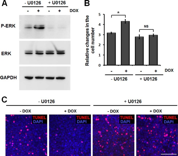
FIGURE 9.
Inhibition of ERK activation abrogates myocilin effects on cell proliferation and survival. A, myocilin-expressing Tet-On HEK293 cells were pretreated with 10 μm U0126 or DMSO for 2 h and further incubated in the medium containing or lacking 1 μg/ml DOX for 48 h. Cell lysates were immunoblotted with anti-phospho-ERK (P-ERK; 1:500 dilution), total ERK (1:1,000 dilution), and anti-GAPDH (1:2,000 dilution) antibodies. B, equal numbers of cells (1 × 10⁵ cells/well) were plated into 6-well plates and incubated as in A. Relative changes in the cell number after incubation in the indicated conditions are shown. Error bars, S.D. of triplicate cultures. NS, non-significant; *, p < 0.05. C, Tet-On HEK293 cells were pretreated with 10 μm U0126 or DMSO for 2 h and then incubated in the medium containing or lacking 1 μg/ml DOX for 24 h. Thereafter, the cells were further incubated in serum-free medium containing 1 μg/ml DOX for 72 h. Apoptotic cells (red fluorescence) were identified as by the TUNEL assay. Similar results were obtained in three independent experiments. The results of a typical experiment are shown. Scale bar, 100 μm.

