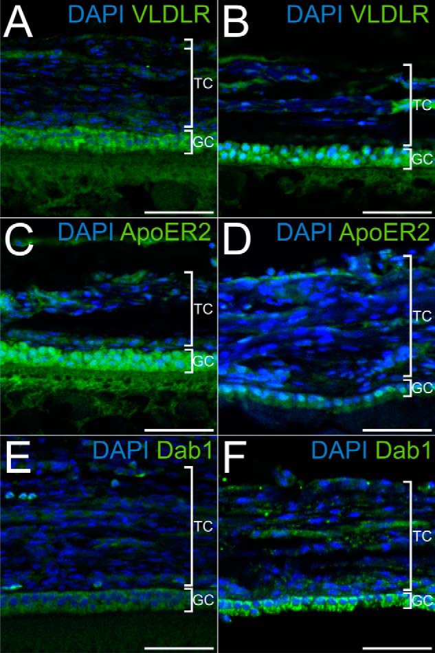
FIGURE 3.

Localization of the components of the Reelin signaling pathway in the chicken follicle. A, C, and E, microtome sections of 5-μm thickness from paraffin-embedded lwf were used to localize the expression of VLDLR (A) with a polyclonal rabbit anti-mouse VLDLR antibody (α187), of ApoER2 (C) with a polyclonal rabbit anti-mouse ApoER2 antibody (α186), and of Dab1 (E) with a rabbit polyclonal anti-mouse Dab1 (D4) antibody. B, D, and F, microtome sections of 5-μm thickness from F6 follicles were used to detect VLDLR, ApoER2, and Dab1 with the same antibodies as in A, C, and E, respectively. In follicles of both developmental stages, VLDLR, ApoER2, and Dab1 are expressed in granulosa cells. TC, theca cells; GC, granulosa cells. Scale bars = 50 μm.
