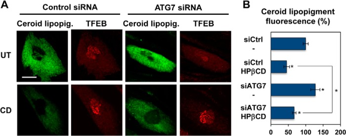
FIGURE 7.

HPβCD-induced activation of autophagy is reduced upon genetic inhibition of the autophagic flux. A, confocal microscopy analysis of ceroid lipopigment (green) and TFEB (red) in LINCL fibroblasts treated with control siRNA or ATG7 siRNA and with 1 mm HPβCD, evaluated by detecting green autofluorescence and binding of an anti-TFEB antibody, respectively. The scale bar is 20 μm. UT, untreated. B, quantification of ceroid lipopigment autofluorescence in LINCL fibroblasts treated as described in A. Representative fields containing ∼50 cells were analyzed. siCtrl, control siRNA; siTFEB, TFEB siRNA. Data are reported as the mean ± S.D. *, p < 0.01.
