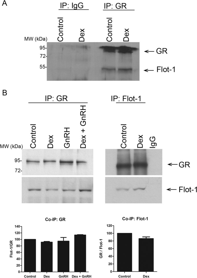
FIGURE 4.

Co-immunoprecipitation (IP) shows that GR and Flot-1 interact ligand independently. A, LβT2 cells were incubated in serum-free medium for 2 h before the addition of 100 nm Dex for 30 min. 400 μl of cell lysates were incubated with GR antibody followed by precipitation with Protein A/G beads. The samples were loaded on an 8% SDS-PAGE gel, transferred onto a nitrocellulose membrane, and probed separately with anti-GR- and anti-Flot-1-specific antibodies. B, as in A, except that cells were also stimulated with 100 nm GnRH and a combination of both Dex plus GnRH, and in the right panel equal amounts of cell lysates were incubated with a rabbit anti-Flot-1 or nonspecific IgG antibody. The top panel shows a single representative Western blot, and the graph shows the combined results of three independent experiments where vehicle (Control) was set to 100%.
