
FIGURE 1.
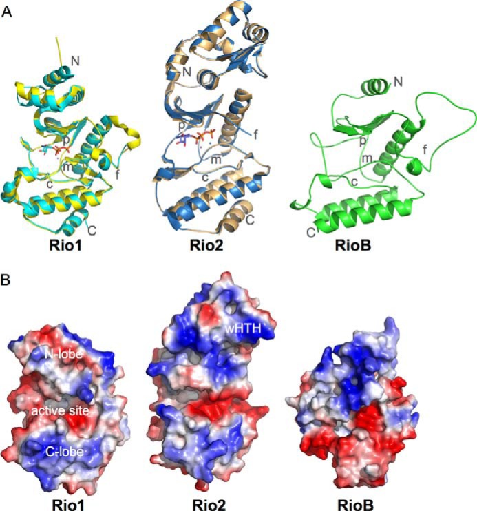
Structure and electrostatic surfaces for prokaryotic RIO kinases. A, the crystal structures of A. fulgidus Rio1 (cyan; Protein Data Bank ID 1ZP9) and Rio2 (blue; ID 1ZAO) bound to ATP superposed on their structures in the absence of nucleotide (yellow and orange, respectively; ID 1ZTF and 1TQI) and a homology model for Tetrasphaera elongata RioB (UniProt N0DXF0). The model was created using MODELLER (23, 24). The N and C termini and the C-, F-, P-, and M-loops are indicated. All structure figures were generated using PyMOL (25). B, electrostatic surfaces for the structures in A in an equivalent orientation. Surfaces were generated using PyMOL (25).