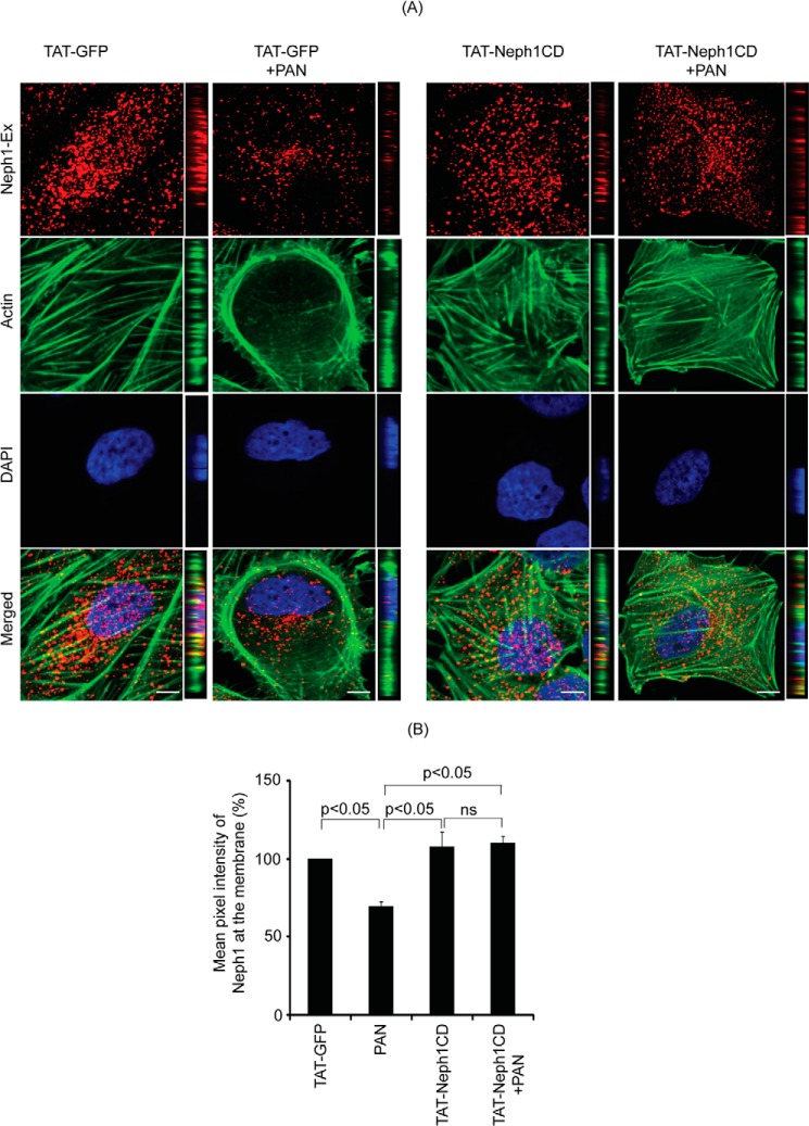
FIGURE 4.
Transduction of TAT-Neph1CD prevents PAN-induced loss of Neph1 from podocyte cell membrane. A, transduced cells were analyzed by immunofluorescence using antibodies directed against actin and the extracellular domain of Neph1 (Neph1-Ex). Confocal images were collected at ×60 magnification and presented after deconvolution in XY and XZ orientation. B, analysis of mean pixel intensity at the membrane of transduced cells suggests that PAN treatment significantly reduced Neph1 localization at the membrane (p < 0.05) in control cells (TAT-GFP+PAN), whereas the cells transduced with TAT-Neph1CD maintained a robust localization of Neph1 at the podocyte cell membrane and were unaffected by treatment with PAN. ns, nonsignificant. Scale bar, A, 10 μm.

