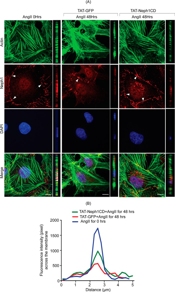
FIGURE 7.
TAT-Neph1CD-transduced podocytes are resistant to cytoskeletal damage induced by Ang II. A and B, TAT-GFP- and TAT-Neph1CD-transduced podocytes were treated with Ang II for the indicated time periods (0–48 h) and immunostained with phalloidin (Alexa 488) and Neph1 (Alexa 594) and mounted with DAPI containing mounting media. Confocal imaging revealed that Ang II treatment resulted in changes to the actin cytoskeleton (where increased stress fibers were noted) along with loss of Neph1 at the cell-cell junctions (A). Notably, the transduction of TAT-Neph1CD prevented these changes (see Fig. 5A). Confocal images were collected at ×60 magnification and constructed after deconvolution in XY and XZ orientation. B, quantitation using pixel intensity profile further demonstrated that similar to PAN, the loss of Neph1 at the cell-cell junctions was minimal in TAT-Neph1CD transduced cells treated with Ang II when compared with the TAT-GFP transduced cells treated with Ang II (p < 0.05). Scale bar, 10 μm (A).

