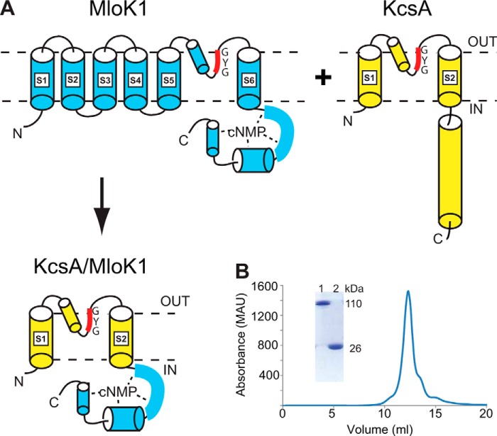
FIGURE 1.
KcsA/MloK1 chimera's design, expression, and purification. A, diagram detailing the expected arrangement of the chimera based on the structures of KcsA (yellow) and MloK1 (blue). The CNBD of MloK1 is directly attached to the bottom of the S6 helix of KcsA. Dashed lines represent the membrane. B, size exclusion chromatography profile of the chimera shows a single peak at a size corresponding to a tetrameric channel in a detergent micelle. Inset shows a portion of an SDS-acrylamide protein gel of unheated tetrameric chimera (lane 1) and heated monomeric chimera (lane 2).

