FIGURE 2.
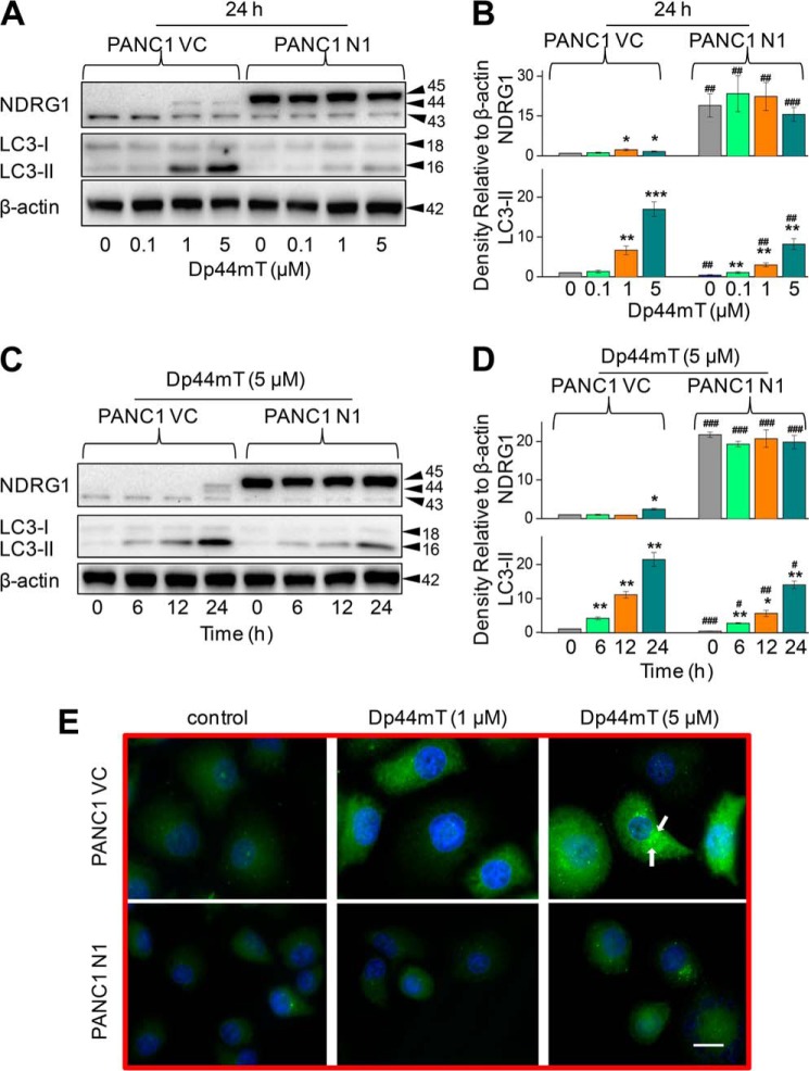
NDRG1 overexpression decreases the classical marker of autophagy, LC3-II, and reduces the number of LC3-II-containing autophagosomes. PANC1 cells were incubated with the iron chelator Dp44mT to examine the effects of NDRG1 overexpression in these cells transfected with the empty vector (PANC1 VC) or NDRG1-containing vector (PANC1 N1). Western blotting was performed to investigate the alteration in expression of LC3-I/II and NDRG1. A, effect of a 24-h, 37 °C incubation of cells with Dp44mT (0.1–5 μm) on NDRG1 and LC3-I/II expression. B, densitometric analysis (arbitrary units) of the results in A. C, effect of incubation time (6–24 h at 37 °C) with 5 μm Dp44mT on NDRG1 and LC3-I/II expression in PANC1 VC and PANC1 N1 cells. D, densitometric analysis (arbitrary units) of the results in C. E, immunofluorescence studies with anti-LC3 antibody in PANC1 VC and N1 cells after a 24-h, 37 °C incubation with control media or this media containing Dp44mT (1 or 5 μm). Scale, 20 μm. The Western analysis and immunofluorescence images shown are typical of three experiments. Densitometric analysis in B and D is the mean ± S.E. (three experiments) normalized to β-actin. *, p < 0.05; **, p < 0.01; ***, p < 0.001 versus their respective controls; #, p < 0.05; ##, p < 0.01; ###, p < 0.001 versus their corresponding treatments in PANC1 VC cells.

