FIGURE 7.
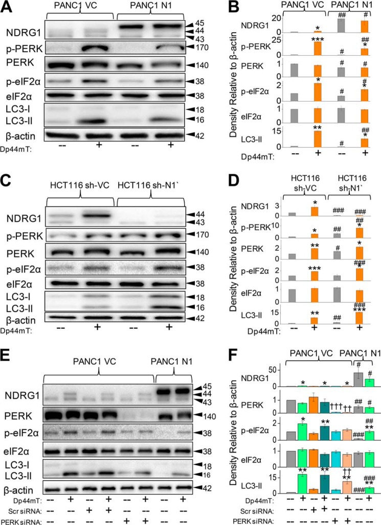
PERK/eIF2α is involved in the NDRG1-mediated suppression of the autophagic pathway. A, PANC1 VC and N1 cells were incubated for 24 h at 37 °C in the presence or absence of Dp44mT (5 μm) to examine its effects on the levels of NDRG1, p-PERK, total PERK, p-eIF2α, total eIF2α, LC3-I, and LC3-II. B, densitometric analysis (arbitrary units) of the blots in A. C, HCT116 sh-VC and sh-N1 cells were incubated for 24 h at 37 °C in the presence or absence of Dp44mT (5 μm) to examine its effects on the levels of NDRG1, p-PERK, total PERK, p-eIF2α, total eIF2α, LC3-I, and LC3-II. D, densitometric analysis (arbitrary units) of the blots in C. E, PANC1 VC cells were preincubated for 48 h at 37 °C with either scrambled (Scr) or PERK siRNA. Cells were then subsequently incubated with Dp44mT (5 μm) for 24 h at 37 °C, and Western analysis performed to investigate levels of NDRG1, total PERK, p-eIF2α, total eIF2α, LC3-I, and LC3-II. F, densitometric analysis (arbitrary units) of the blots in E. The Western analysis shown is typical of three experiments. Densitometric analysis is the mean ± S.E. (three experiments) normalized to β-actin: *, p < 0.05; **, p < 0.01; ***, p < 0.001 versus their respective controls. #, p < 0.05; ##, p < 0.01; ###, p < 0.001 versus their corresponding treatments in PANC1 VC for A or E or HCT116 sh-VC for C cells; ††, p < 0.01; †††, p < 0.001 versus their corresponding treatments in cells treated with the scrambled control.

