FIGURE 8.

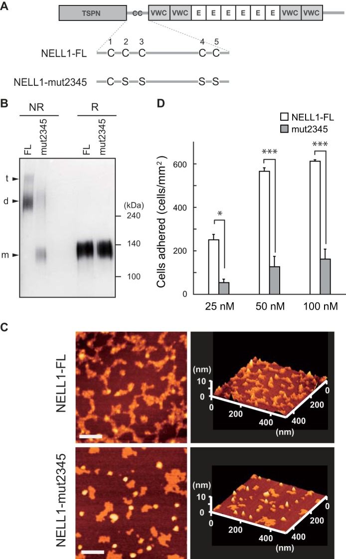
A quadruple cysteine-to-serine mutant of NELL1 shows reduced cell adhesion activity. A, schematic of intact (NELL1-FL) and quadruple cysteine-to-serine substitution mutant (NELL1-mut2345) NELL1s. E, EGF-like repeat. B, the intact and mutant NELL1 proteins were expressed in 293-F cells, and the purified proteins were resolved by SDS-PAGE under non-reducing (NR) or reducing (R) conditions and visualized by silver staining. The bands assumed to be monomers (m), dimers (d), and trimers (t) are indicated at the left. Molecular weight markers are indicated at the right. C, atomic force microscopy images of NELL1-FL and NELL1-mut2345. Samples were adsorbed onto a mica chip, dried, and then scanned in dynamic force microscopy mode. Scale bars = 100 nm. D, NELL1-FL and NELL1-mut2345 were coated onto 96-well plates at a concentration of 25, 50, or 100 nm, and MC3T3-E1 cells were allowed to adhere for 1.5 h at 37 °C before adhered cells in the wells were processed for counting. Each value represents the mean ± S.E. of triplicate results. *, p < 0.05; ***, p < 0.001 (Student's t test).
