FIGURE 1.
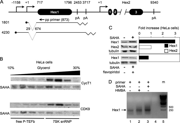
The HEXIM1 and HEXIM2 genes, HEXIM1 transcripts, and their responses to SAHA and HMBA. A, schematic of the HEXIM1 (Hex1) and HEXIM 2 genes and HEXIM1 transcripts. The HEXIM1 gene measures 4875 bp and contains only one exon (Hex1), but two transcripts are observed (the transcription start sites are labeled using ●). 645 bp are spliced out of both transcripts before this exon. Numbers start with the major transcription start sites of the HEXIM1 (this study) and HEXIM2 genes. The HEXIM1 gene also contains two polyadenylation (pA) sites, which are indicated by zigzag lines. The HEXIM2 gene measures 9340 bp and contains three exons. The annealing position of the promoter-proximal (pp) primer for 5′ RACE was at position 873 from the transcription start site (equivalent to position 157 of exon 1 in the HEXIM1 gene). B, SAHA releases free P-TEFb from the 7SK snRNP. Cell lysates of HeLa cells treated with DMSO or SAHA (5 μm for 90 min) were subjected to a 10–30% glycerol gradient sedimentation (the black bar indicates increasing glycerol concentrations). Anti-CycT1 or anti-CDK9 antibodies detected subunits of P-TEFb in each fraction by western blotting. C, only HEXIM1, but not HEXIM2, is inducible by SAHA in HeLa cells. The + and −signs indicate the presence and absence of SAHA (top and bottom panels) and flavopiridol (bottom panel), respectively. HeLa cells were treated with SAHA for 48 h. Western blot analyses were quantified, and data are presented as fold increase by SAHA. Only HEXIM1 was induced 3-fold by SAHA (top western blot and black bar), and its induction was abrogated by flavopiridol (bottom western blot and black bar). α-Tubulin was used as the loading control. These western blot analyses are representative of three independent experiments that yielded the same results. D, only the previously unannotated proximal HEXIM1 promoter is inducible by SAHA and HBMA in HeLa cells (lanes 1-3). The pp primer is presented in A. Lane 1 represents resting HeLa cells. Lane 2 is from HeLa cells treated with SAHA. Lane 3 is from HeLa cells treated with HMBA. Lane 4 is a blank control. Lane 5 is the DNA marker, defined as m in panel D.

