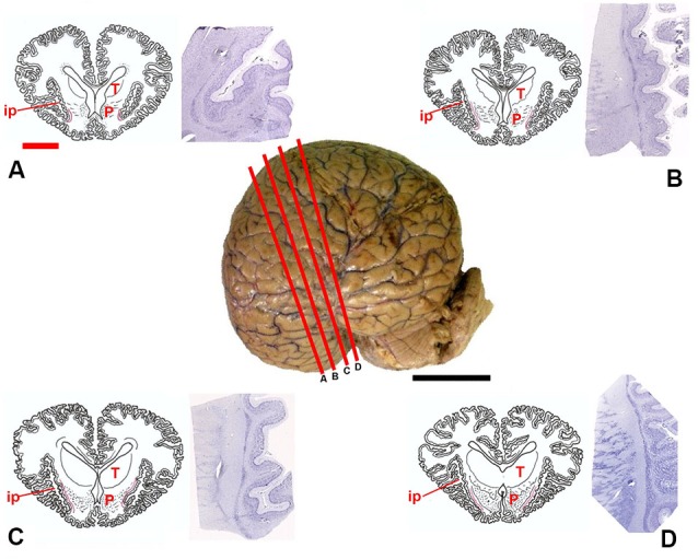
Figure 1.

Position of the claustrum in Tursiops truncatus. Center, lateral photograph of the adult brain; the orange bars (A–D) approximate the levels of subsequent sections. The drawings (A–D) are based on composite renderings of coronal sections of the dolphin brain, sided by Nissl preparations obtained approximately at the corresponding levels. In the drawings the claustrum is represented by a thin red line. Note the reduction of the endopiriform cortex ventral to the basal ganglia; P, putamen; T, thalamus; ip, insular pocket. Black scale bar (lateral photograph of the dolphin brain) = 5 cm; orange scale bar (drawings) = 2 cm.
