Figure 4.
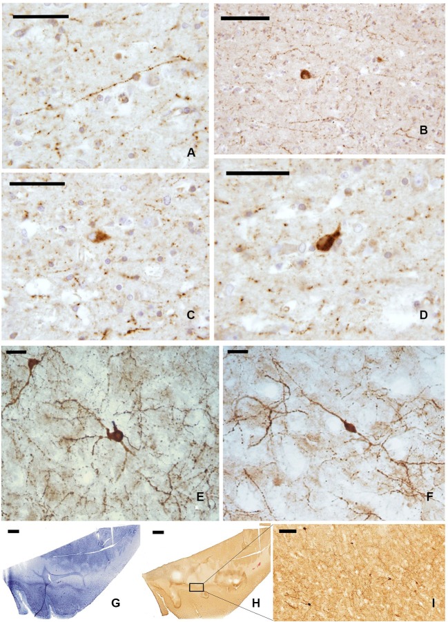
NPY-ir fibers (A, B) and neurons (C, D) in paraffin-embedded sections of the claustrum; (E, F) NPY positive neurons in frozen sections; (G–I) localization of NPY-ir neurons in whole sections ((G) Nissl stain, (H–I) immunocytochemistry); I represent an enlargement of the black rectangle in H. Scale bars = A, C, D = 50 μm; B, I = 100 μm; E, F = 20 μm;. G, H = 2 mm.

