FIGURE 1.
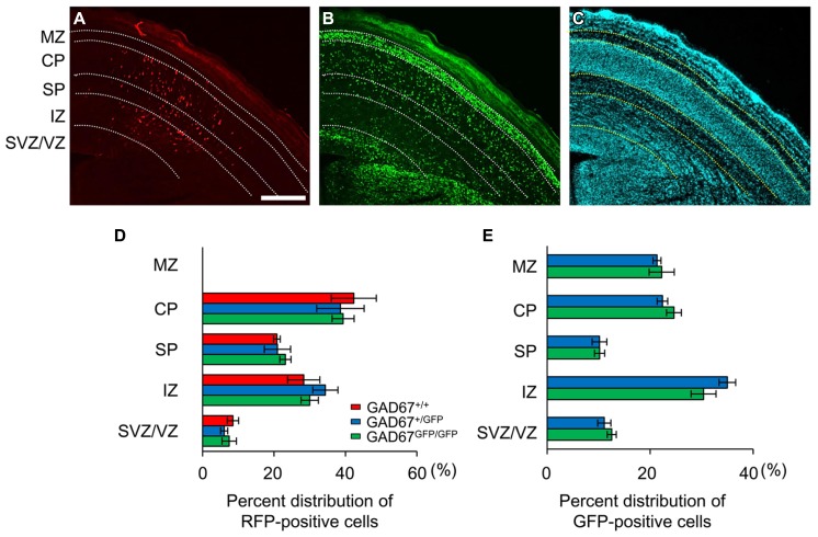
Distribution patterns of radially migrating neurons and tangentially migrating GABAergic neurons in the cerebral cortex at E17.5. (A) Distribution of radially migrating neurons expressing mRFP (red) in a coronal section of the neocortex of a GAD67+/GFP fetus. (B) Distribution of GABAergic neurons expressing GFP (green) in the same section. (C) The section was counterstained with DAPI. The dotted lines in (A–C) indicate the boundaries of the proliferative and migratory zones. The scale bar represents 200 μm. MZ, marginal zone; CP, cortical plate; SP, subplate; IZ, intermediate zone; SVZ, subventricular zone; VZ, ventricular zone. (D) Percent distributions of RFP-positive cells in the CP, SP, IZ, and SVZ/VZ averaged over five GAD67+/+ fetuses, four GAD67+/GFP fetuses, and five GAD67GFP/GFP fetuses. (E) Percent distributions of GFP-positive cells in the MZ, CP/SP, IZ, and SVZ/VZ (GAD67+/+: n = 4; GAD67+/GFP: n = 5). Error bars indicate the mean ± SEM.

